Department of ultrasound diagnostics
The first ultrasound studies in GOKOD began in 1989 with the physician Regina Moiseevna Karpenko on the only at that time apparatus "Aloka SSD-630".
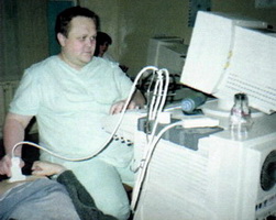 The number of studies barely reached two hundred per year and consisted mainly of studies of the thyroid gland in children.
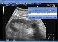 At present, up to 34 thousand people pass through the department, and the spectrum of studies includes almost all methods and techniques known to modern ultrasound diagnostics, in addition, the vascularization of tumor tissues and parenchymatous organs is assessed, practically the whole range of diagnostic and therapeutic minimally invasive manipulations is under control Ultrasound. In March 2000, diagnostic trepan-biopsy of peripheral lung tumors and formations of the anterior and posterior mediastinum was developed and widely used. With the acquisition of modern kits, it became possible to widely apply drainage procedures, the application of percutaneous nephropyelostomies, and drainage of the bile ducts both for palliative purposes, and in terms of preoperative and pre-radial preparations.
In recent years, our department has been equipped with new ultrasound scanners of high and expert class, due to which we were able to do three-dimensional reconstruction of volumetric structures and a more reliable definition of the spread of tumor invasion, an in-depth diagnosis of prostate pathology.
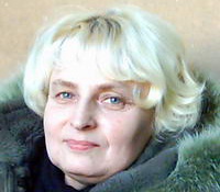
The quality of the work of the department directly depends on the high qualification of the doctors of the department - Regina Moiseevna Karpenko, Irina Vitalievna Burmistrova, Marina Vladlenovna Murycheva.
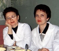 Unfortunately, for various reasons, our department was left by such high-class specialists as Valentina Vladimirovna Bondarenko and Oksana Belyan, who made a significant contribution to the development of ultrasound diagnostics of the dispensary. But time does not stand still and in recent years the branch has replenished with young, but experienced and promising specialists - Murashko Konstantin Leonidovich and Maloletnikova Khristina Mikhailovna.
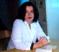 In November 2015, the department, dispensary and all ultrasound diagnostics of the Republic of Belarus suffered a heavy blow - the permanent head of our department, one of the founders and chairman of the society of specialists in ultrasonic diagnostics of the Gomel region, the luminary of ultrasonic diagnostics, Oleg Ivanovich Anikeev, died suddenly. Oleg Ivanovich's contribution to medicine is huge, it is difficult to overestimate. We lost a friend, a teacher and a doctor with a capital letter. The loss is irreplaceable, but experience and colossal knowledge Oleg Ivanovich handed over to his students, who also maintain a high level of ultrasound diagnosis in the dispensary.
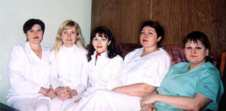 The sisters of the branch are of great help in the work. Irina Vinogradova, Elena Bazhenova, Diana Sutorma, Efremenko Antonina Alexandrovna. The quality of their work could be envied by the Kremlin clinic, and the speed of printing - the best stenographers. And the work of the senior nurse of our department Mukomelo Svetlana Viktorovna has always been an example for imitation and admiration.
From the left to the right: Ovchinnikova Svetlana (department chief's assistant), Oksana Dyatlova (doctor), Irma Vinogradova, Kublitskaya Larisa (nurses), Anisimova Lena (sister-hostess)
Branch phones:
8 (0232) 49 19 02, 8 (0232) 49 18 90Address: 246000 Republic of Belarus, Gomel, ul.
Medical, 2. Regional clinical oncology dispensary.
Possibilities of separation of ultrasonic diagnostics GKOD
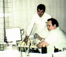
Understanding the ultrasonic diagnostics GOKOD has been the flagship of the ultrasonic diagnostics of Gomel and the region for more than 15 years, residents of all corners of the Republic of Belarus, and, more recently, of the near abroad aspire to explore in different ways. While single, but not rare, patients from the United States, Britain, Germany and Israel.
Our advisory opinions are in most cases the final diagnosis, and they concern not only oncological pathology, but affect all areas of practical medicine.
In addition to covering the entire range of currently known echoscopic studies, we are also leaders in the field of interventional sonography, when under ultrasound control, after a preliminary anesthesia, a puncture of the pathological formation takes place, either to take material for various types of further research, or to conduct medical procedures.
Similarly, the formation of the thyroid gland, mammary gland, abdominal organs, small pelvis, inorganic formations, soft tissues and much more, that is all that can be seen with the help of ultrasound, is investigated.
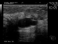 To illustrate and confirm this, those who wish can view the video, where "disappearance" of the mammary gland and the filling of its lumen with 96% alcohol for the complete cure of this pathology occurs on your eyes.
The procedure takes several minutes, and the patient no longer needs to prepare for a surgical operation.The method is confirmed by the existence of multiple large cysts of the mammary glands, the restriction on one procedure is caused only by the permissible amount of alcohol administered at a time, because a certain amount of it is absorbed through the walls of the cyst and causes not a sad state known to most of our patients :).
Cysts of the kidneys and the thyroid gland are treated in a similar way. In some cases (which are determined jointly by an endocrinologist and a doctor of ultrasound) with the help of alcoholization, it is possible to perform non-operative treatment of non-cystic, and nodular pathology of the thyroid gland.
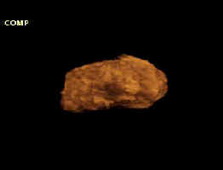
Widely, together with urologists, we are engaged in the pathology of the prostate gland. The latest achievements in hardware support allow a complete scanning of the organ with the formation of a three-dimensional volume image with subsequent detailed study, differentiation of inflammatory and tumor pathology.
Those interested can familiarize themselves with the 3D (3D) ultrasound reconstruction of the prostate gland and make sure that it is not only in essence, but also in the form of a second heart of a man :).
Significant help in establishing the correct diagnosis is provided by the presence of intracavity sensors with high spatial resolution and equipped with a sensitive color Doppler, which allows to identify formations that in the usual case could not be determined before the operation by any other existing method.
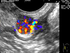 To the left - a node of 2 cm of the small pelvis in a woman with a large number of pathological vessels, to the right - a node of 8 mm of the prostate (dark zone with blood vessels in the left lobe, in the right lobe blood supply is normal).
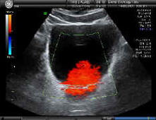
High-quality dopplerography, in addition to its main purpose, also makes it possible to assess the functional abilities of the non-vascular nature.
You have a unique opportunity to see the manifestation of the kidneys.
Those flames that blaze in the bladder are nothing but the result of urine ejection from both ureters during their normal functioning. Do not be afraid, this fire does not burn!
No intravenous injection, no contrasts - and the result is obvious ...
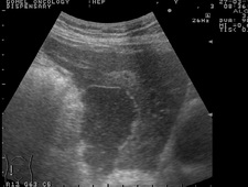
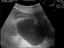
Of the non-tumorous diseases that can be treated well in our dispensary without surgery, and by puncture and drainage under the supervision of ultrasound, abscesses (abscesses), bruises and other developed fluid formations.
As examples of this pathology, it is possible to demonstrate a postoperative liver abscess (left) and a pseudocyst of the pancreas (right), which can be completely cured by this method.Click to enlarge the picture.
Combined use of techniques of high-resolution ultrasound color Doppler mapping and biopsy under ultrasound guidance to help deal with this non-cancer disorders such as chronic hepatitis, including viral and autoimmune etiology, cirrhosis of the liver, various metabolic disorders, nephritis and nephrosis, chronic pancreatitis, thyroiditis and Much, much more.
Even this cursory review is enough to show our diagnostic capabilities. It is easier to find a method that we do not apply in our activity, than vice versa.
The incentive to improve is constantly increasing need for new research, new approaches to the diagnosis and treatment of many of our patients.
Witnesses of our achievements are more than 26 thousand patients, which we examine per year and which we carry out more than 70 thousand studies.
The experience of minimally invasive interventions is estimated at more than 800 procedures per year.
An important indicator of trust is the fact that 10 years on the basis of our dispensary there have been meetings of the Society of Ultrasound Diagnostic Doctors of the Gomel Region.


