Magnetic resonance imaging (MRI)
Magnetic resonance imaging (MRI) is a safe, painless, exceptionally accurate and reliable modern research method. This is the peak of the modern diagnostic pyramid.
MRT-tomograph is a tunnel with a circular magnetic coil. Inside the tunnel for a short period of time creates an electromagnetic field of high voltage. Under the influence of this field, the hydrogen nuclei that are in the water molecules (the human body consists mainly of water molecules) are turned and aligned in the direction of the field. After the field is turned off, the protons return to their original state. The difference in the energies of the two proton states is released in the form of photons, which are caught by the scanner of the tomograph. The essence of the method is to analyze the behavior of the nuclei of hydrogen atoms in the human body in a constant magnetic field of high intensity. Healthy cells and diseased cells generate different signals, and the tomograph fixes this. This is the principle of the MRI scanner.
Thus, the patient is not exposed to radiation during the study.
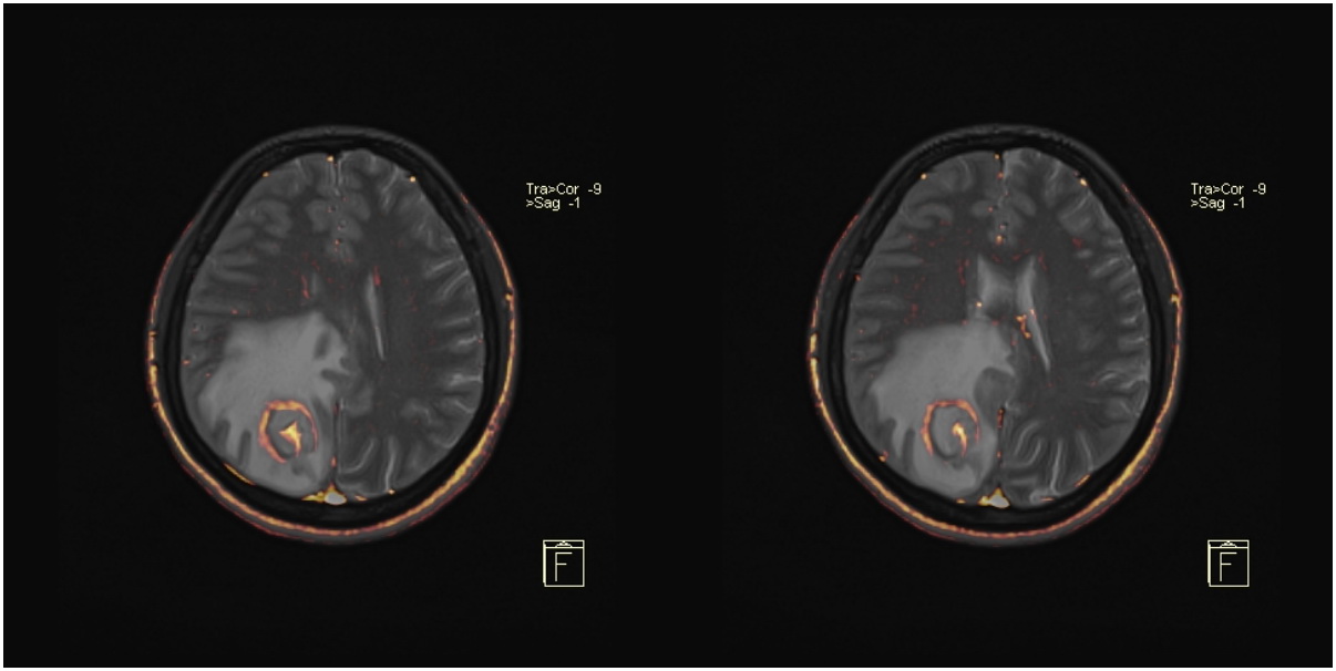
Magnetic resonance imaging in many cases makes it possible to detect changes in tissues and organs that can not be detected by other methods. MRI is most informative in the early detection and diagnosis of tumors, but this method has no analogues in various diseases of non-tumoral nature. MRI allows you to visualize the brain and spinal cord in detail, as well as assess the structure of other internal organs with high quality.
The development of MRI was rapid, and if earlier this method was used as a refining diagnosis, now it is increasingly used for the initial diagnosis and preventive examinations.
Possibilities of magnetic resonance imaging in GKODC
One of the most important characteristics of any tomograph is the intensity of the magnetic field, measured in Tesla. The higher the given value, the more accurately the results of the study are obtained.
Our institution is equipped with a high-field magnetic resonance tomograph Magnetom Avanto from Siemens with a field strength of 1.5 Tesla (Tesla).
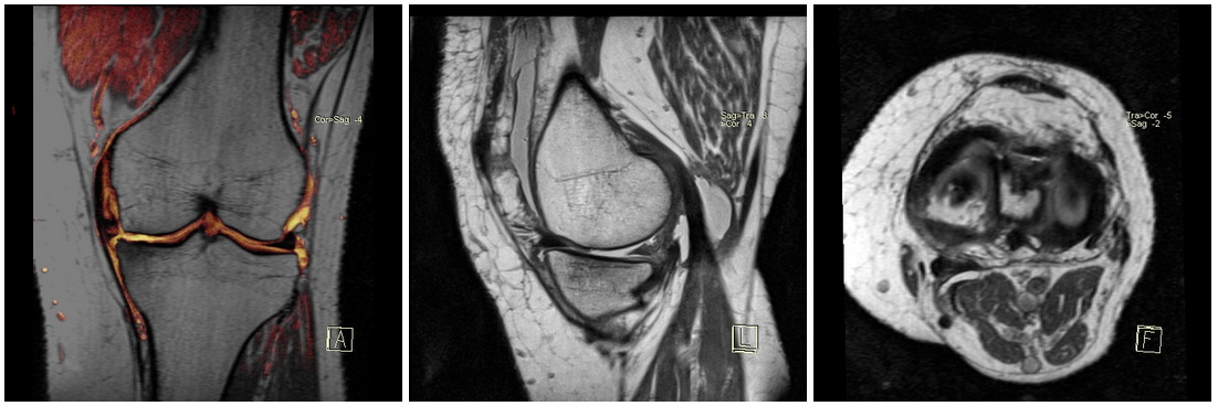
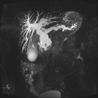
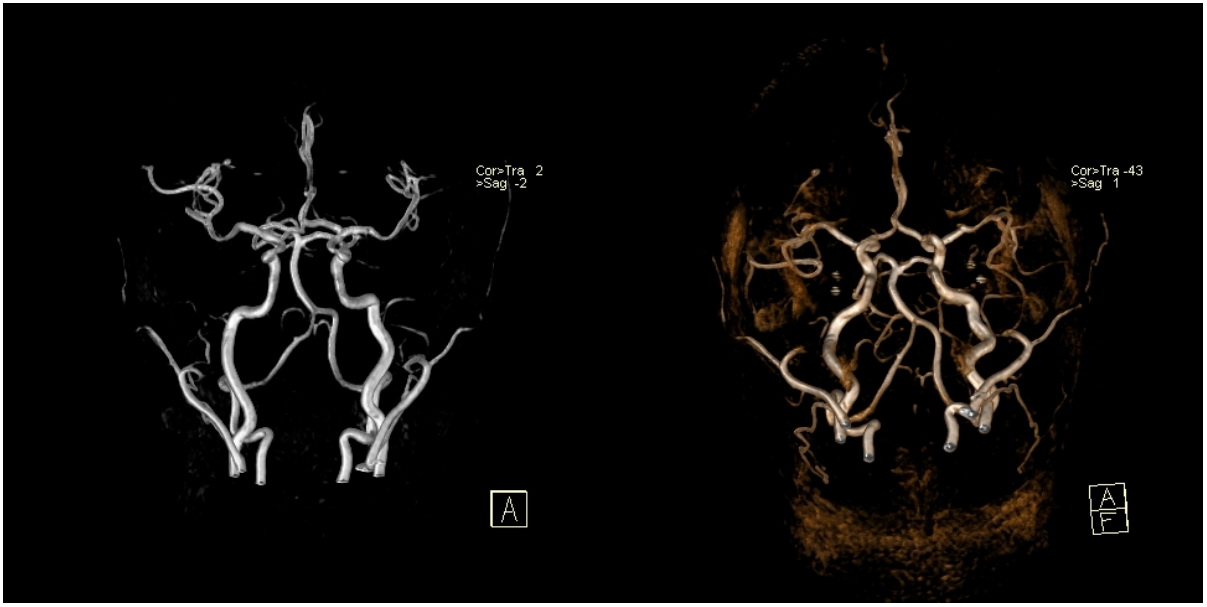
The tomograph makes it possible to obtain both planar and bulk anatomical images of the investigated region by the method of magnetic resonance.
Magnetic resonance imaging is used in the diagnosis:
- Diseases of the brain and spinal cord;
- Oncological diseases;
- Diseases of the genitourinary system;
- Diseases of the breast;
- Diseases of bones and joints.
In our institution it is possible to perform special methods of MRI diagnostics:
- MRI with the "diffusion" protocol. Allows you to obtain accurate information about the movement of water molecules through the cell membranes.
- MRI with contrast. This type of MRI allows you to obtain data on the movement of blood in the tumor. It is carried out with an intravenous injection of a special substance that improves the visualization of the scanned object. The most informative for differential diagnosis of tumor diseases.
- Spectroscopy. It gives an opportunity to receive information about chemical cellular activity.
- Perfusion MRI. It is carried out to obtain information on the blood flow at the capillary level.
Information for patients
Absolute contraindications to MRI
- Artificial heart rate drivers (can go into asynchronous mode of operation under the influence of a magnetic field);
- Intracranial ferromagnetic haemostatic clasps of the cerebral vessels (with displacement may occur vessel damage and bleeding);
- Aortic clips;
- Electrodes
- Ferromagnetic metal implants;
- Metal structures in the anatomical area to be investigated (metal plates, distractors, etc.);
- Periorbital ferromagnetic foreign bodies (if damaged, damage to the eyeball may occur);
- Ferromagnetic or electronic implants of the middle ear;
- Pronounced claustrophobia;
- Restriction of the weight of the patient (not more than 125 kg)
Relative contraindications to MRI
- Moderate claustrophobia;
- Epilepsy, schizophrenia;
- Pregnancy (especially in the first trimester);
- Decompensated heart failure;
- Prosthetic heart valves;
- Hemostatic clips of other localization;
- Implanted neurostimulants or leads;
- Insulin pump;
- Extremely serious condition of the patient;
- The need for physiological monitoring;
- The impossibility for the patient to remain immobile during the examination.
Most medical devices are conditionally compatible with MRI. This means that the examination of patients with established stents, intravascular coils, filters, prosthetic heart valves can be carried out in the presence of clinical indications in agreement with a specialist in radiation diagnosis on the basis of information from the manufacturer on the characteristics of the metal from which the installed device is manufactured.
The presence of metal teeth, tantalum brackets on the sternum is not a contraindication to the study, although it may reduce image quality.
Preparation
Most of the MR studies do not require special training, except for the study of the abdominal organs, which is recommended to be performed on an empty stomach. In this case, for 6-8 hours before the start of the study you need to refrain from eating. This preparation is necessary to obtain the most clear images.
Wear comfortable clothes without metal fragments and take with you a change of shoes. Decoration and costume jewelry should be left at home. Before the study is not recommended to use cosmetics containing metal particles, and metal-containing ointments.
If you have metal implants (except dental crowns), a pacemaker, artificial heart valves, endoprostheses, any foreign bodies, or if you are afraid of enclosed spaces, please inform your doctor. For any of the above cases, the specialist will give you advice on the possibility of an MRI study.
Sometimes, full and effective research is performed with local or complete anesthesia, especially for young children who can not remain immobile during the procedure.
How is the examination conducted
During the study, you will be constantly monitored by medical personnel. An MRI scan can take from 15 minutes to an hour, depending on the volume and complexity of the study.
In practice, it looks like this: a patient on a special table is placed inside a tunnel of a tomograph in which a magnetic field is created - for a person completely safe. Within a few minutes, a scan is performed, and then the computer processes the received data and displays the image on the monitor screen.
After the end of the study, the results are analyzed by a radiologist. This takes a certain time, so the results of the study and the protocol of description will be passed on to your doctor. In case of emergency medical indications and in consultation with a doctor, results can be obtained within 1 hour after the examination.
Original images are copied to your CD-R drive.
In the offices of MSCT and MRI are doctors of the first and highest qualification category who have been trained in the MNIOR them. AP Gerzena, at the St. Petersburg State University, Bristol General Hospital (England).
Our modern equipment allows you to quickly and accurately diagnose. Our staff is highly qualified specialists with extensive experience in the field of disease diagnosis.
In the department, X-ray studies are carried out to ensure the II level of visualization:
- All types of traditional X-ray studies, including radiography, fluoroscopy, linear tomography;
- Special studies (mammography, fistulography, retrograde contrasting of the biliary tract, excretory and retrograde urography, etc.);
- X-ray computed tomography
The main directions of diagnostic research:
- Diagnosis of diseases of the organs of the gastrointestinal tract (esophagus, stomach, small and large intestine). The department introduces the technique of primary double contrasting of the digestive tract with the use of an original contrast agent;
- Diagnosis of breast diseases;
- Studies of the osteoarticular apparatus;
- Organs of the chest cavity;
- Urinary tract;
- Bile ducts;
contact number
Head of the division Mikhail Mikhailovich
Gapeenko :
8 (0232) 49 18 83 , 8 (0232) 49 15 02 , 8 (0232) 49 19 34 .
Also mentally to sort out the questions that have arisen, you can e-mail mikgfer@tut.by